Confocal microscopy: pinhole, PSF product and optical sectioning
A wide field fluorescence microscope images every plane of a thick sample at once: the focal plane sharply, all others as a blurred haze. Minsky’s confocal microscope (patent 1957) illuminates one point at a time with a focused spot and detects through a pinhole in the image plane. Light from above or below the focus is defocused at the pinhole and mostly blocked. Two things follow:
the effective PSF is the product of the excitation PSF and the detection PSF (convolved with the pinhole), slightly narrower than the wide field PSF,
a thin fluorescent plane gives a signal that falls off with its distance from focus: optical sectioning, which wide field imaging cannot do at all.
Function ( |
Purpose |
|---|---|
|
3D amplitude PSF from the pupil (uniform or aplanatic) by angular spectrum propagation |
|
pinhole sizes in Airy units, 1 AU = \(1.22\lambda/\mathrm{NA}\) |
|
the fluorescence confocal PSF for any pinhole |
|
signal of a thin fluorescent plane vs. defocus (sectioning) |
|
axial response of a reflection confocal to a mirror |
Sections: 0. the setup (drawn with pyoptics.sketch); 1. the 3D wide field PSF of an oil objective; 2. the confocal
PSF; 3. the pinhole: resolution, sectioning and signal; 4. optical
sectioning, fluorescence and reflection; 5. an xz section through a thick
sample, wide field vs. confocal.
The pupil and Abbe’s picture are from notebook 52; transmitted light methods for transparent samples in 53. The 3D PSF is scalar here; the vectorial high NA focus (Richards-Wolf) is in notebook 02, and PSF, MTF and the Strehl ratio in 17. Conventions: \(e^{i(\mathbf{k}\cdot\mathbf{r}-\omega t)}\), lengths in micrometres in object space, \(z\) along the axis in the immersion medium.
import time

import numpy as np
import matplotlib.pyplot as plt
from scipy.ndimage import gaussian_filter

from pyoptics import microscopy as mic
from pyoptics.utils import grid2d

plt.rcParams.update({"figure.dpi": 80, "image.origin": "lower", "font.size": 11})
t_start = time.time()

n_oil = 1.518
NA = 1.4                    # oil immersion objective
wl_exc, wl_em = 0.488, 0.520   # excitation (argon/diode laser) and emission (GFP), um
AU = mic.airy_unit(wl_em, NA)
N, dx = 256, 0.04           # the confocal PSF reaches 4 NA/lambda: dx <= lambda/(8 NA) = 0.044 um
L = N*dx
x, y, X, Y = grid2d(-L/2, L/2, -L/2, L/2, N)
z = np.linspace(-2, 2, 81)
dz = z[1] - z[0]
alpha = np.arcsin(NA/n_oil)
print(f"aperture angle {np.degrees(alpha):.1f}°, 1 AU = {AU:.3f} um (emission, object space)")


def fwhm(coord, profile):
    # full width at half maximum of a single peak, with linear interpolation
    p = profile/np.max(profile)
    i0 = np.argmax(p)
    above = p >= 0.5
    left = i0
    while left > 0 and above[left - 1]:
        left -= 1
    right = i0
    while right < len(p) - 1 and above[right + 1]:
        right += 1
    xl = np.interp(0.5, [p[left - 1], p[left]], [coord[left - 1], coord[left]])
    xr = np.interp(0.5, [p[right + 1], p[right]], [coord[right + 1], coord[right]])
    return xr - xl


h_exc = mic.psf_3d(x, y, z, wl_exc, NA, n=n_oil, apodization="aplanatic")
h_det = mic.psf_3d(x, y, z, wl_em, NA, n=n_oil, apodization="aplanatic")
I_wf = np.abs(h_det)**2                # wide field fluorescence: the emission PSF
c = N//2
iz0 = len(z)//2
aperture angle 67.3°, 1 AU = 0.453 um (emission, object space)
0. The laser scanning confocal microscope
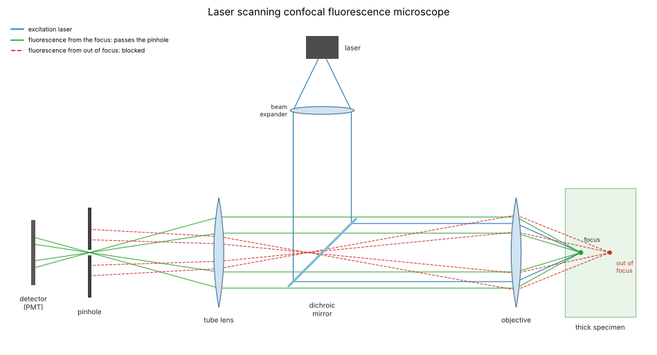
The layout, unfolded except for the dichroic mirror: the laser beam is expanded to fill the objective’s pupil and focused to a diffraction limited spot in the sample (blue). Fluorophores there emit at a longer wavelength (green); the emission returns through the objective, passes the dichroic mirror, which reflects the laser line and transmits the fluorescence, and is focused by the tube lens onto the pinhole, a field plane conjugate to the focus. Light from a fluorophore deeper in the sample (red, dashed) comes to a focus in front of the pinhole and is spread over a disk at the pinhole, so the pinhole blocks most of it. Scan mirrors in a pupil plane (omitted here) move the spot across the sample, and the image is built up point by point from the detector signal.
from pyoptics import sketch

fig, ax = plt.subplots(figsize=(12, 6.6))
sketch.confocal_microscope(ax, title="Laser scanning confocal fluorescence microscope")
plt.tight_layout()
1. The 3D wide field PSF
psf_3d() propagates the pupil to every plane with the exact
\(k_z = \sqrt{n^2k_0^2 - k_\perp^2}\) (a scalar Debye integral); the
aplanatic option weights the pupil with \((n k_0/k_z)^{1/2}\), the amplitude
of an objective that obeys the sine condition. The classic estimates for
the widths (FWHM) of the intensity PSF are
the latter a non-paraxial version of \(2n\lambda/\mathrm{NA}^2\). On the axis
the uniform pupil gives exactly
\(|h(0,z)| = |\langle e^{i(k_z - nk_0)z}\rangle_\mathrm{pupil}|\), which
mirror_response(z/2) evaluates (a mirror doubles the defocus), and in
the paraxial limit \(\mathrm{sinc}^2(u/4\pi)\) with \(u = k_0\mathrm{NA}^2z/n\).
The right panel shows the key fact for sectioning: the light in each plane \(\int I\,dx\,dy\) is the same everywhere. Defocus spreads the light but does not remove it, so a wide field microscope sees a uniform fluorescent layer equally bright at any focus.
h_unif = mic.psf_3d(x, y, z, wl_em, NA, n=n_oil)
fig, axes = plt.subplots(1, 3, figsize=(20, 5.2), gridspec_kw=dict(width_ratios=[1.2, 1, 1]))
ax = axes[0]
xz = I_wf[:, c, :].T
im = ax.imshow(np.log10(xz/xz.max() + 1e-4), extent=(z[0], z[-1], x[0], x[-1]), aspect="auto",
               cmap="magma", vmin=-3, vmax=0)
plt.colorbar(im, ax=ax, fraction=0.046, pad=0.04, label="log10 I")
ax.set_xlabel("z (µm)"); ax.set_ylabel("x (µm)"); ax.set_ylim(-1.5, 1.5)
ax.set_title(f"wide field PSF, x-z section (NA {NA} oil, {wl_em*1000:.0f} nm)")
ax.plot([0, 2], [0, 2*np.tan(alpha)], "w:", lw=1)
ax.plot([0, 2], [0, -2*np.tan(alpha)], "w:", lw=1)

ax = axes[1]
u = 2*np.pi/wl_em*NA**2*z/n_oil
ax.plot(z, I_wf[:, c, c], color="tab:blue", lw=2, label="aplanatic pupil")
ax.plot(z, np.abs(h_unif[:, c, c])**2, color="tab:orange", lw=2, label="uniform pupil")
ax.plot(z[::3], mic.mirror_response(z[::3]/2, wl_em, NA, n=n_oil), "ko", mfc="none", ms=4,
        label="uniform, mirror_response(z/2)")
ax.plot(z, np.sinc(u/(4*np.pi))**2, "k--", lw=1, label="paraxial sinc²(u/4π)")
ax.set_xlabel("z (µm)"); ax.set_ylabel("on-axis intensity"); ax.legend(fontsize=9)
ax.set_title("axial profile")

ax = axes[2]
E = np.sum(I_wf, axis=(1, 2))
ax.plot(z, E/E[iz0], lw=2, label=r"$\int I\,dx\,dy$ (wide field)")
ax.set_ylim(0, 1.2); ax.set_xlabel("z (µm)"); ax.set_ylabel("light per plane")
ax.set_title("no optical sectioning in wide field"); ax.legend(fontsize=9)
plt.tight_layout()

dx_wf, dz_wf = fwhm(x, I_wf[iz0, c]), fwhm(z, I_wf[:, c, c])
print(f"wide field FWHM: lateral {dx_wf*1000:.0f} nm (0.51 lambda/NA = {0.51*wl_em/NA*1000:.0f} nm), "
      f"axial {dz_wf*1000:.0f} nm (0.88 lambda/(n - sqrt(n^2 - NA^2)) = "
      f"{0.88*wl_em/(n_oil - np.sqrt(n_oil**2 - NA**2))*1000:.0f} nm)")
wide field FWHM: lateral 186 nm (0.51 lambda/NA = 189 nm), axial 503 nm (0.88 lambda/(n - sqrt(n^2 - NA^2)) = 491 nm)
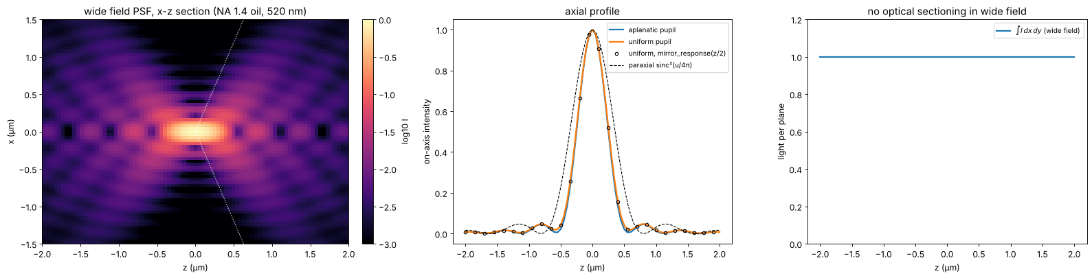
The aplanatic weighting puts more weight on the pupil edge and makes the focus slightly narrower and the axial profile asymmetric; the uniform pupil agrees with the exact on-axis integral, while the paraxial formula overestimates the axial extent at this NA by about 40 %.
2. The confocal PSF
The laser spot excites a fluorophore at \(\mathbf{r}\) with \(|h_\mathrm{exc}(\mathbf{r})|^2\). Its emitted light forms the spot \(|h_\mathrm{det}|^2\) in the image plane, centred at the (back projected) position of the fluorophore, and the pinhole \(D\) passes the fraction \((|h_\mathrm{det}|^2 \otimes D)(\mathbf{r})\). Fluorescence is incoherent, so the PSF of the scanned image is the product
For a point pinhole it is \(|h_\mathrm{exc}|^2|h_\mathrm{det}|^2\): for equal wavelengths the square of the wide field PSF, \(\sqrt{2}\) times narrower for a Gaussian-like core (FWHM \(\approx 0.37\lambda/\mathrm{NA}\) laterally, \(0.64\lambda/(n - \sqrt{n^2 - \mathrm{NA}^2})\) axially), and the side lobes are suppressed. Open pinholes approach the excitation PSF.
pinholes = {"point pinhole": None, "1 AU": AU}
psf_c = {name: mic.confocal_psf(h_exc, h_det, x, y, d) for name, d in pinholes.items()}
lam_mean = np.sqrt(wl_exc*wl_em)
fig, axes = plt.subplots(2, 3, figsize=(20, 9.6))
for j, (name, P) in enumerate([("wide field", I_wf)] + list(psf_c.items())):
    ax = axes[0, j]
    xz = P[:, c, :].T
    im = ax.imshow(xz/xz.max(), extent=(z[0], z[-1], x[0], x[-1]), aspect="auto", cmap="magma",
                   vmin=0, vmax=1)
    plt.colorbar(im, ax=ax, fraction=0.046, pad=0.04)
    ax.set_xlabel("z (µm)"); ax.set_ylabel("x (µm)"); ax.set_ylim(-1, 1); ax.set_xlim(-1.5, 1.5)
    ax.set_title(f"{name}: x-z section (linear)")
ax = axes[1, 0]
for name, P in [("wide field", I_wf)] + list(psf_c.items()):
    ax.plot(x, P[iz0, c]/P[iz0, c].max(), lw=2, label=f"{name}: FWHM {fwhm(x, P[iz0, c])*1000:.0f} nm")
ax.set_xlim(-0.8, 0.8); ax.set_xlabel("x (µm)"); ax.set_ylabel("normalized intensity")
ax.set_title("lateral profiles"); ax.legend(fontsize=9)
ax = axes[1, 1]
for name, P in [("wide field", I_wf)] + list(psf_c.items()):
    ax.plot(z, P[:, c, c]/P[:, c, c].max(), lw=2, label=f"{name}: FWHM {fwhm(z, P[:, c, c])*1000:.0f} nm")
ax.set_xlabel("z (µm)"); ax.set_ylabel("normalized intensity")
ax.set_title("axial profiles"); ax.legend(fontsize=9)
ax = axes[1, 2]
for name, P in [("wide field", I_wf)] + list(psf_c.items()):
    row = P[iz0, c]/P[iz0, c].max()
    ax.semilogy(x, row, lw=1.5, label=name)
ax.set_ylim(1e-6, 1.5); ax.set_xlim(0, 1.5); ax.set_xlabel("x (µm)")
ax.set_title("side lobes (log scale)"); ax.legend(fontsize=9)
plt.tight_layout()
print(f"point pinhole: lateral {fwhm(x, psf_c['point pinhole'][iz0, c])*1000:.0f} nm "
      f"(0.37 lambda/NA = {0.37*lam_mean/NA*1000:.0f} nm), axial "
      f"{fwhm(z, psf_c['point pinhole'][:, c, c])*1000:.0f} nm "
      f"(0.64 lambda/(n - sqrt(n^2 - NA^2)) = {0.64*lam_mean/(n_oil - np.sqrt(n_oil**2 - NA**2))*1000:.0f} nm)")
point pinhole: lateral 129 nm (0.37 lambda/NA = 133 nm), axial 351 nm (0.64 lambda/(n - sqrt(n^2 - NA^2)) = 346 nm)
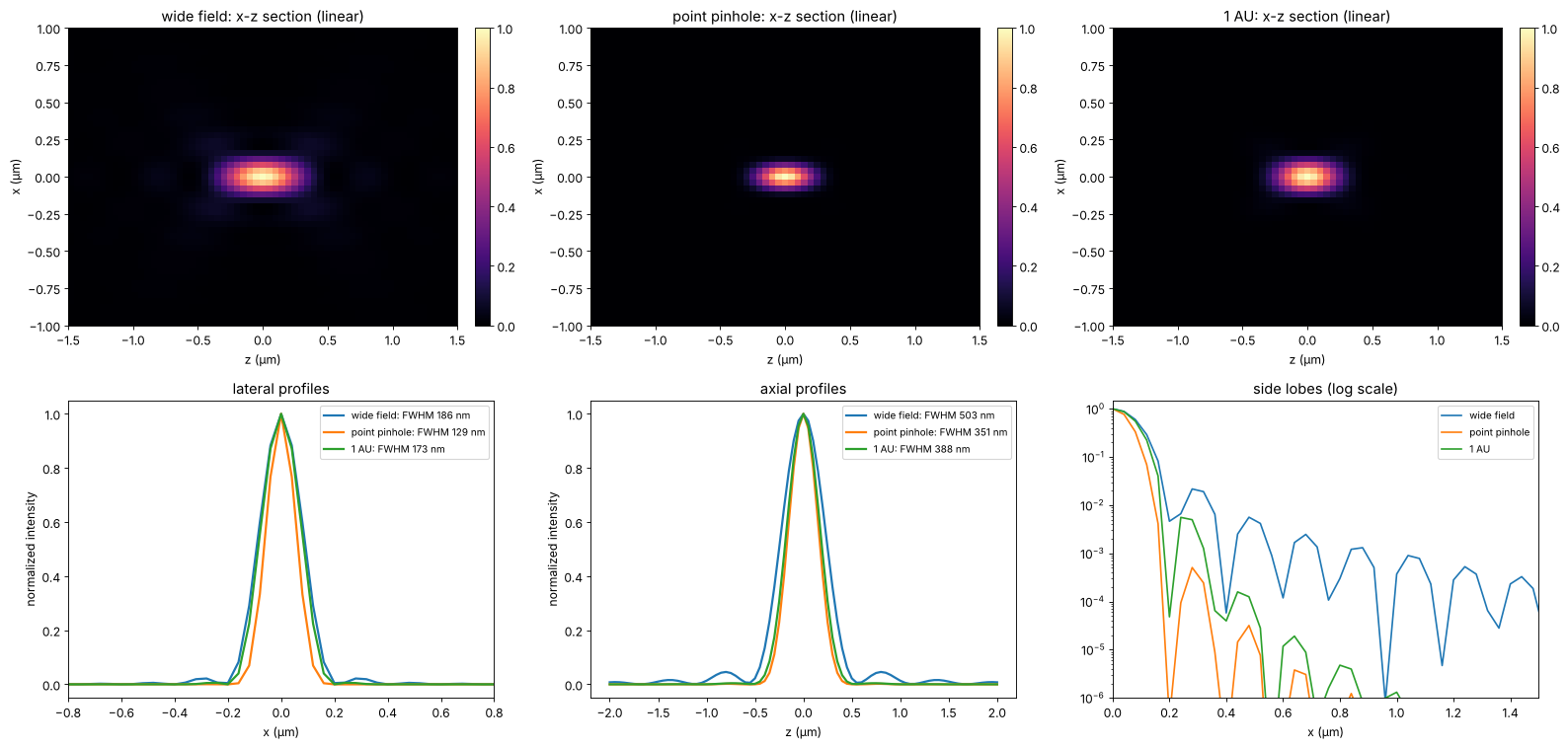
(The estimates use the geometric mean of the two wavelengths.) The point pinhole is a theoretical limit: it collects no light. In practice the pinhole is set to about 1 AU, where the lateral resolution is close to wide field and the gain of confocal microscopy lies almost entirely in the sectioning.
3. The pinhole: resolution, sectioning and signal
How large may the pinhole be? The plot follows the lateral and axial FWHM of the PSF of a point and the detected signal from a point in focus, for pinholes from 0 to 3 AU. The full lateral gain needs pinholes below about 0.25 AU, which pass only a quarter of the light; by 1 AU it is mostly gone. The axial width of the PSF degrades more slowly. Beyond 1 AU the signal grows only slowly (the paraxial Airy disk holds 84 % of the light, the aplanatic high NA PSF here somewhat less), so 1 AU is the usual compromise.
d_au = np.array([0.0, 0.1, 0.25, 0.5, 0.75, 1.0, 1.5, 2.0, 3.0])
lat, ax_w, sig = [], [], []
for d in d_au:
    P = mic.confocal_psf(h_exc, h_det, x, y, d*AU if d > 0 else None)
    lat.append(fwhm(x, P[iz0, c]))
    ax_w.append(fwhm(z, P[:, c, c]))
    sig.append(P[iz0, c, c])
lat, ax_w, sig = map(np.array, (lat, ax_w, sig))
# signal relative to the full Airy pattern (an infinitely large pinhole)
sig_rel = sig/(np.abs(h_exc[iz0, c, c])**2*np.sum(np.abs(h_det[iz0])**2))
fig, axes = plt.subplots(1, 2, figsize=(16, 5))
ax = axes[0]
ax.plot(d_au, lat/dx_wf, "o-", lw=2, label="lateral FWHM / wide field")
ax.plot(d_au, ax_w/dz_wf, "s-", lw=2, label="axial FWHM / wide field")
ax.axvline(1.0, color="0.7", ls=":")
ax.set_xlabel("pinhole diameter (AU)"); ax.set_ylabel("relative width")
ax.set_title("PSF widths vs. pinhole"); ax.legend(fontsize=9); ax.set_ylim(0.5, 1.15)
ax = axes[1]
# a point pinhole collects nothing; pinholes below one pixel are not resolved on this grid
ok = d_au >= 0.25
ax.plot(np.r_[0, d_au[ok]], np.r_[0, sig_rel[ok]], "o-", lw=2, color="tab:green")
ax.axhline(0.838, color="0.7", ls=":", label="84 % (light in the Airy disk)")
ax.axvline(1.0, color="0.7", ls=":")
ax.set_xlabel("pinhole diameter (AU)"); ax.set_ylabel("detected fraction of the emitted light")
ax.set_title("signal from a point in focus"); ax.legend(fontsize=9)
plt.tight_layout()
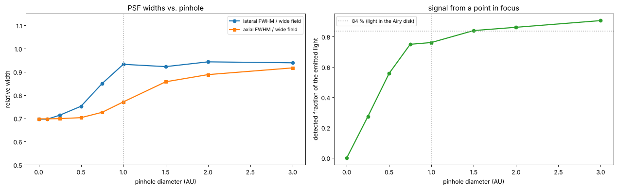
The axial FWHM of the PSF of a point (a bead) stays within the wide field value even for large pinholes, because the excitation spot alone is already confined; what changes dramatically is the response to extended objects, the subject of the next section.
4. Optical sectioning
The standard test of sectioning is a thin uniform fluorescent layer moved
through the focus. Its signal is the integral of the PSF over each plane
(plane_response()): constant for wide field, falling off for confocal.
Far from focus the defocused detection spot of radius \(\approx z\tan\alpha\)
overfills the pinhole, so the detected fraction falls like
\(1/z^2\); the excitation light per plane is constant. Here a larger window
(20 µm) is used so that the defocused spots do not wrap around, and the
planes are computed one by one.
A reflection confocal microscope seen by a plane mirror (point
pinhole) is coherent: the reflected field is the pupil with twice the
defocus, and the detected intensity is
\(|\langle e^{2i(k_z - nk_0)z}\rangle|^2\) (mirror_response()), in the
paraxial limit \(\mathrm{sinc}^2(u/2\pi)\), the first zero at
\(z = n\lambda/\mathrm{NA}^2\). This is the principle of confocal
profilometers for surface topography.
N2, dx2 = 512, 0.04
L2 = N2*dx2
x2, y2, _, _ = grid2d(-L2/2, L2/2, -L2/2, L2/2, N2)
z2 = np.linspace(-4, 4, 81)
resp = {"wide field": [], "confocal 0.5 AU": [], "confocal 1 AU": [], "confocal 2 AU": []}
for zi in z2:
    he = mic.psf_3d(x2, y2, zi, wl_exc, NA, n=n_oil, apodization="aplanatic")
    hd = mic.psf_3d(x2, y2, zi, wl_em, NA, n=n_oil, apodization="aplanatic")
    resp["wide field"].append(np.sum(np.abs(hd)**2))
    for name, d in [("confocal 0.5 AU", 0.5), ("confocal 1 AU", 1.0), ("confocal 2 AU", 2.0)]:
        resp[name].append(np.sum(mic.confocal_psf(he, hd, x2, y2, d*AU)))
fig, axes = plt.subplots(1, 2, figsize=(16, 5.2))
ax = axes[0]
widths = {}
for name, r in resp.items():
    r = np.array(r)/np.max(r)
    if name != "wide field":
        widths[name] = fwhm(z2, r)
    ax.plot(z2, r, lw=2, label=name + (f" (FWHM {widths[name]:.2f} µm)" if name in widths else ""))
zm = np.linspace(-4, 4, 801)
I_m = mic.mirror_response(zm, wl_exc, NA, n=n_oil)
ax.plot(zm, I_m, "k--", lw=1, label=f"reflection, mirror (FWHM {fwhm(zm, I_m):.2f} µm)")
ax.set_xlabel("z (µm)"); ax.set_ylabel("normalized signal"); ax.set_ylim(0, 1.1)
ax.set_title("thin fluorescent plane moved through the focus"); ax.legend(fontsize=9)
ax = axes[1]
for name, r in resp.items():
    r = np.array(r)/np.max(r)
    sel = z2 > 0
    ax.loglog(z2[sel], r[sel], "o-", ms=3, lw=1.5, label=name)
zz = np.array([1.5, 4])
ax.loglog(zz, 0.35*(zz/1.5)**-2, "k:", label=r"$\propto 1/z^2$")
ax.set_xlabel("z (µm)"); ax.set_ylabel("normalized signal")
ax.set_title("far from focus"); ax.legend(fontsize=9)
plt.tight_layout()
for name, w in widths.items():
    print(f"{name}: plane response FWHM {w:.2f} um")
confocal 0.5 AU: plane response FWHM 0.39 um
confocal 1 AU: plane response FWHM 0.50 um
confocal 2 AU: plane response FWHM 0.81 um
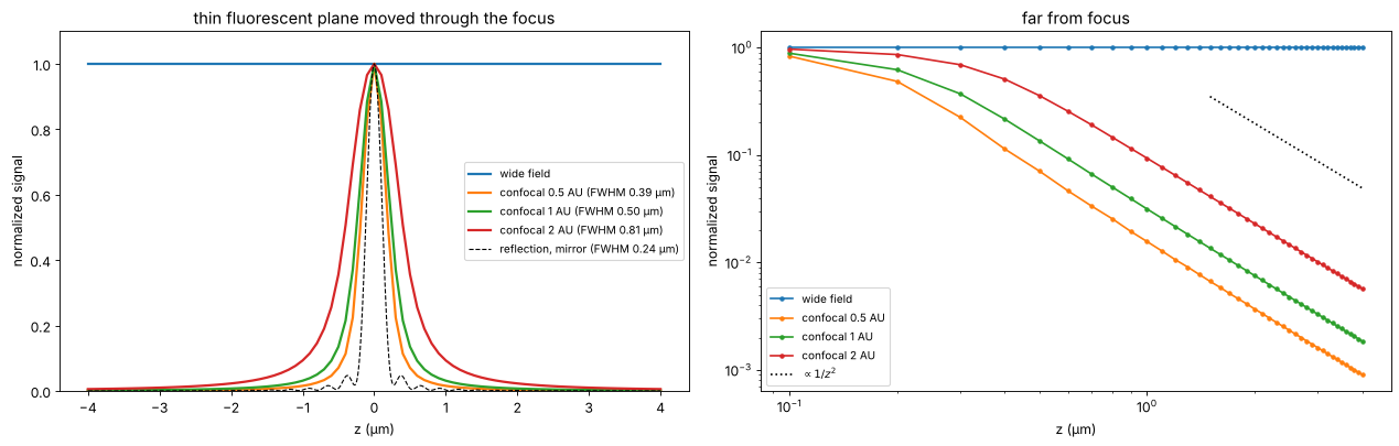
The sectioning width grows with the pinhole: about 0.5 µm at 1 AU for this objective, 1.6 times that at 2 AU. The reflection response with its point pinhole is narrower still (0.24 µm, while the paraxial \(0.89\,n\lambda/\mathrm{NA}^2\) would give 0.34 µm), and its side lobes are the zeros of the coherent sinc.
5. An x-z section through a thick sample
The final test: a fluorescent “cell” with a membrane, a nucleus envelope and filaments, all extended along \(y\) (so that the 3D convolution reduces to a 2D convolution in \(x\) and \(z\) with the PSF integrated over \(y\)). The wide field image (an x-z stack of focus positions) drowns the section in out-of-focus light and stretches everything along \(z\); the confocal image at 1 AU shows a clean section. The PSFs are computed on a 10 × 10 µm x-z window and the sample is placed in its centre.
z3 = np.arange(-64, 64)*0.08            # 10.24 um, same span as x
psf_wf_xz, psf_cf_xz = [], []
for zi in z3:
    he = mic.psf_3d(x, y, zi, wl_exc, NA, n=n_oil, apodization="aplanatic")
    hd = mic.psf_3d(x, y, zi, wl_em, NA, n=n_oil, apodization="aplanatic")
    psf_wf_xz.append(np.sum(np.abs(hd)**2, axis=0))
    psf_cf_xz.append(np.sum(mic.confocal_psf(he, hd, x, y, AU), axis=0))
psf_wf_xz, psf_cf_xz = np.array(psf_wf_xz), np.array(psf_cf_xz)   # (z, x)

ZZ, XX = np.meshgrid(z3, x, indexing="ij")
r_ell = np.hypot(XX/3.6, ZZ/2.2)
# thin shells as smooth rings, thicker than the z sampling of 0.08 um
sample = 1.0*np.exp(-((r_ell - 1)/0.05)**2)                                        # membrane
sample += 0.6*np.exp(-((np.hypot((XX - 0.6)/1.4, (ZZ + 0.2)/1.0) - 1)/0.08)**2)   # nuclear envelope
for x0, z0 in [(-2.2, 0.8), (-1.6, -1.1), (1.9, 1.2), (2.6, -0.4), (0.2, 1.5), (-0.4, -1.5)]:
    sample += 3*np.exp(-((XX - x0)**2 + (ZZ - z0)**2)/(2*0.05**2))           # filaments along y
sample += 0.25*(r_ell < 1)                                  # cytoplasmic background
sample = gaussian_filter(sample, 1.0)                       # no stair steps from the grid


def image(psf):
    return np.real(np.fft.ifft2(np.fft.fft2(sample)*np.fft.fft2(np.fft.ifftshift(psf/psf.sum()))))


img_wf, img_cf = image(psf_wf_xz), image(psf_cf_xz)
fig, axes = plt.subplots(1, 3, figsize=(20, 5.6))
ext = (x[0], x[-1], z3[0], z3[-1])
for ax, img, title in [(axes[0], sample, "sample (fluorophore density), x-z"),
                       (axes[1], img_wf, "wide field x-z stack"),
                       (axes[2], img_cf, "confocal x-z stack, 1 AU")]:
    im = ax.imshow(img/img.max(), extent=ext, cmap="gray", vmin=0, vmax=1, aspect="equal")
    plt.colorbar(im, ax=ax, fraction=0.046, pad=0.04)
    ax.set_xlim(-4.5, 4.5); ax.set_ylim(-3.2, 3.2)
    ax.set_xlabel("x (µm)"); ax.set_ylabel("z (µm)"); ax.set_title(title)
plt.tight_layout()
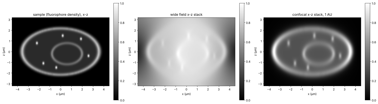
Summary
The wide field PSF spreads the light of each plane over a growing disk but keeps it: a uniform layer looks the same at every focus, there is no optical sectioning.
The confocal PSF is the product of excitation PSF and detection PSF convolved with the pinhole (
confocal_psf()). A point pinhole narrows the PSF by about \(\sqrt{2}\) and suppresses the side lobes, but collects no light; at the usual 1 AU the lateral resolution is close to wide field.The real gain is optical sectioning: the signal of a fluorescent plane falls off as \(1/z^2\) (
plane_response()), with a width of about 0.5 µm at 1 AU and NA 1.4, growing with the pinhole above 1 AU.A reflection confocal (coherent, mirror) gives the response \(|\langle e^{2i(k_z - nk_0)z}\rangle|^2\) (
mirror_response()), with the paraxial width \(0.89\,n\lambda/\mathrm{NA}^2\) (narrower at high NA).
Image scanning microscopy (Airyscan and similar) replaces the pinhole by a small detector array and reassigns the pixels to recover the \(\sqrt{2}\) without losing light; it builds on exactly the PSF product computed here.
print(f"run time {time.time() - t_start:.0f} s")
run time 31 s
References
M. Minsky, “Microscopy apparatus”, US patent 3,013,467 (1961, filed 1957); M. Minsky, “Memoir on inventing the confocal scanning microscope”, Scanning 10, 128-138 (1988).
T. Wilson and C. Sheppard, Theory and Practice of Scanning Optical Microscopy, Academic Press (1984).
C. J. R. Sheppard and T. Wilson, “Depth of field in the scanning microscope”, Opt. Lett. 3, 115-117 (1978).
T. Wilson (ed.), Confocal Microscopy, Academic Press (1990).
J. B. Pawley (ed.), Handbook of Biological Confocal Microscopy, 3rd ed., Springer (2006).
B. Richards and E. Wolf, “Electromagnetic diffraction in optical systems II. Structure of the image field in an aplanatic system”, Proc. R. Soc. Lond. A 253, 358-379 (1959).
C. B. Müller and J. Enderlein, “Image scanning microscopy”, Phys. Rev. Lett. 104, 198101 (2010).
J. Mertz, Introduction to Optical Microscopy, 2nd ed., Cambridge University Press (2019).